Vías de tratamiento para la artrosis de rodilla
Written by:The doctor. of Barutell, a renowned specialist in Pain, Anesthesiology and Resuscitation Unit, explains the available treatment routes for osteoarthritis of the knee. The doctor was founder of the Pain Clinic of the Hospital Vall d'Hebrón, first Clinic of the Pain of Catalonia and second of Spain. He is currently co-director of the Hospital El Pilar Pain Clinic.
The knee joint is the largest synovial joint in the human body, allowing for the extension and flexion movements of the leg, while ensuring a good stability and resistance to the weight to be supported, as well as sufficient mobility for move it.
It consists of three joints: two femhototibial joints , which transfer body weight to the legs, between the femoral and tibial condyles, and a patellofemoral joint between the patella and the femur.
There are also two menisci , external and internal, between the femur and the knee, which cushion the articular surfaces during movement. The joint is reinforced by extracapsular ligaments that stabilize the hinge movement of the knee (internal lateral, external lateral, patellar, oblique popliteal and arcuate popliteus) and intracapsular ligaments that maintain the contact of the articular surfaces during the flexion of the knee (ligament crossed anterior and posterior). All joint surfaces uncoated with articular cartilage are lined by the synovial membrane, with several synovial sacs.
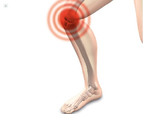
Knee osteoarthritis: what it is and how it manifests
Osteoarthritis of the knee is a chronic disease of very high prevalence in adults older than 45 years, being more prevalent in women than in men.
It is characterized by mechanical pain , functional deterioration, joint instability and periarticular muscle weakness. Initially insidious, with episodes of pain that increase in frequency and duration. When joint damage is severe in some patients, there is continuous pain. Pain and stiffness are located at the joint level and are not accompanied by general symptomatology. Morning stiffness after rest is usually brief and localized. The mechanisms that can provide pain are: increased intracapsular and intraosseous pressure, subchondral microfractures, involvement of the tendons or bursitis secondary to muscle weakness and injuries of joint structures.
In the exploration one can observe pain in the passive movements and crackling of the articular structures. In advanced osteoarthritis, there is some degree of joint instability and deformity due to the formation of juxtaarticular osteophytes.
How Osteoarthritis of the Knee is Diagnosed and Imaging
Diagnosis is usually as a result of physical examination and the findings of simple radiology although, sometimes, it may happen that a patient has severe pain that is not correlated with the radiological findings, due to the existence of superadded lesions, such as the meniscal rupture. It is also frequent that patients with radiological alterations of osteoarthritis are asymptomatic. Therefore, therapeutic decisions, especially surgical decisions, should always be based on the articular functional stage and the repercussions of pain on the patient's activities and not on imaging tests.
The classic radiological findings of osteoarthritis are: osteophytes, impingement of the joint space, subchondral sclerosis, articular geodas and subluxations. In addition, magnetic resonance imaging (MRI) is currently the technique with the greatest diagnostic capacity in the field of musculoskeletal radiology, as it allows the study of bone, articular and soft tissue. Therefore, the technique of choice can be considered after a conventional radiological study. MRI allows to quantify in a sensitive and reproducible way the loss of cartilage in osteoarthritis .
Treatments available for knee osteoarthritis
The treatment of osteoarthritis of the knee will be individualized by the specialist in Unity of Pain. A non-pharmacological approach that includes patient education, weight management and rehabilitation exercises appropriate to each patient is considered fundamental, especially to strengthen the quadriceps. A wide array of pharmacological agents with anti-inflammatory drugs (NSAIDs) and, ultimately, opioids are also available.
Intraarticular blockade or infiltration of the knee is a very therapeutic modality to be taken into account in the osteoarthritis of this joint. In general, it achieves a great relief of the symptoms, with few side effects, being a complementary or alternative treatment to others. Joint infiltration can be performed with different drugs: corticosteroids, local anesthetics, hyaluronic acid and platelet-rich plasma and growth factors. The following are detailed:
- Application of intra-articular corticosteroids → Requires delayed formulations, the most indicated being triamcinolone acetonide. They exert their effect quickly, are reliable and have few side effects. It is not advisable to repeat four more infiltrations in the same joint and must be separated more than one week between them. However, they are not free of risks such as postinjection pain, tendon rupture, microcrystalline arthritis and joint infection.
- Local anesthetics → They can be used alone but are usually mixed with corticosteroids to reduce the appearance of arthritis by microcrystals , while at the same time producing an early relief of symptoms.
- Hyaluronic acid (HA ) → It is a natural component of the matrix of connective tissue and synovial fluid of the joints. The basis of its use lies in the attempt to improve altered joint biomechanics in osteoarthritis. It exerts a restorative action of viscoelasticity and restorative on the joint lubricating function. We currently have multiple high or low molecular weight preparations in pre-filled syringes of 3 to 5 doses that are given weekly. In addition, there are single dose preparations (for a single injection) with higher doses, which is the one we usually use. They may be effective in improving pain and joint function between 6-8 months although it depends on several factors, such as the age of the patient, severity of the disease and duration of the disease.
- Plasma rich in platelets and growth factors → It is an autologous blood product with a high platelet content, the result of the centrifugation of a blood sample. When injected into the joint, the growth factors derived from platelets are added and the disease is improved.. The articular hyaline cartilage of the knee is an avascular, aneural and allophatous tissue, with scarce possibilities of scarring itself, due to its scarce cellular population. The application of platelet-rich plasma helps repair the damage of this tissue , especially in the earliest stages of the disease.
If all these treatments do not give the expected result you can always resort to joint replacement surgery.