Lateral external ligament
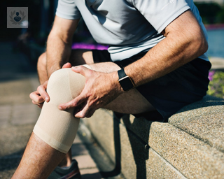
The lateral collateral ligament (LCL) or lateral collateral ligament is one of four major ligaments that support the knee joint. Connects the femur to the tibia: it goes from the top of the tibia in the lower section of the femur. Its function is not to allow lateral mobility of the knee joint stable and maintain the outer side of the joint. The most common injuries occur by pressure or an injury that pushes the knee joint from the inside, which results in stress on the outside. The symptoms of a rupture or tear of the lateral collateral ligament are: swelling of the knee, blocking knee motion, pain or tenderness on the outer side of the knee, the knee gives way or feels like to do when It is active or voltage. You must make an examination of the lateral collateral ligament laxity possible to study this. It involves bending the knee to 25 degrees and putting pressure on the inner surface. They should also take X-rays of the joint, MRI, among other studies. The patient should apply ice to the area, in addition to anti-inflammatory, knee and constantly raise physical activity until the pain and swelling go away.

Lesiones ligamentosas de rodilla: ¿qué son y cómo se tratan?
By Dr. Fernando Ávila España
2024-04-24
El Dr. Fernando Ávila España explica qué son y en qué consisten las lesiones ligamentosas de rodilla, así como suelen producirse y cómo se tratan en la actualidad. De hecho, el Dr. Ávila aplica un innovador sistema en e que ya no utiliza la inmovilización tras la cirugía. See more
Experts in Lateral external ligament
-

Bernardo Prueba
Orthopaedic SurgeryExpert in:
- Dysplasia
- Knee fracture
- Anterior cruciate ligament
- Posterior cruciate ligament
- Lateral external ligament
-

Dr. Vicente Jesús León-Muñoz
Orthopaedic SurgeryExpert in:
- Knee prosthesis
- Hip replacement surgery
- Posterior cruciate ligament
- Anterior cruciate ligament
- Meniscus
- Lateral external ligament
- See all

Instituto de Cirugía Avanzada de Rodilla (ICAR)
Instituto de Cirugía Avanzada de Rodilla (ICAR)
C. Barítono Marcos Redondo, 1. 7ºB.
No existe teléfono en el centro.
By using the telephone number provided by TOP DOCTORS, you automatically agree to let us use your phone number for statistical and commercial purposes. For further information, read our Privacy Policy
Top Doctors
-
Instituto de Cirugía Avanzada de Rodilla (ICAR)
C. Barítono Marcos Redondo, 1. 7ºB., MurciaExpert in:
- Arthroscopic surgery
- Regenerative Medicine
- Trauma
- Child Trauma