Symptoms and Treatments of Retinal Detachment
Written by: Retinal detachment consists of the separation of the r 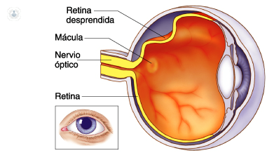 Of the pigment epithelium, which is the underlying tissue. It is caused by the accumulation of liquid between them.
It affects 1 in 10,000 people. Although it can affect at any age, it is more frequent between 40 and 70 years of age, as well as in myopic patients (40% of cases), who have suffered trauma (10% of cases) and those with a family history Of retinal detachment. In 10% of cases both eyes are affected.
Symptoms of retinal detachment
Typical symptoms are: vision of flashes or lights, blurred vision or defects in the visual field referred to as a gray or black curtain by the patient.
The miodesopsias or flies are characterized by being transparent objects of diverse and mobile forms that are interposed in the visual field. They are usually multiple and black, and may be caused by retinal pigment epithelial cells released into the vitreous cavity or bleeding.
The photopsies or luminous flashes are usually repetitive and located on the same sector of the visual field, being able to indicate the presence of vitreoretinal traction. This may lead to suspicion about the presence of a retinal rupture, although they do not always appear as a previous symptom.
Sometimes the patient notices directly the complete or partial loss of visual field , referring to it as a black curtain that descends and covers the vision. This symptom may suggest a retinal detachment.
If the detachment area is very small peripheral or symptoms may be virtually nonexistent.
Any of these symptoms should put us on the alert and is reason to consult with an ophthalmologist. In the case of retinal tear, early treatment may prevent retinal detachment. If it has already occurred, waiting time until surgery is an important risk factor in view of the patient's visual prognosis.

Diagnosis of retinal detachment
The anamnesis gives us valuable information, as well as a very approximate diagnostic orientation. Before any of the symptoms described above should be referred immediately to the ophthalmologist for a complete eye examination. Visual acuity should be analyzed and the fundus of the eye should be explored with complete pupil dilation by indirect ophthalmoscopy with or without scleral indentation.
Clinic
Retinal detachment can be classified in three ways if we consider the mechanism of production of the same: regmatogenic, tractional and exudative.
- Regmatogenous: caused by retinal tear or breakage frequently occurring after a posterior vitreous detachment. This allows the passage of a liquefied vitreous into the subretinal space, which causes separation of the neurosensory retina.
- Tractional: caused by the tensions that produce membranes or the newly formed tissue. They pull the retina and lift it. The main cause is proliferative diabetic retinopathy.
- Exudative : in this case the retina does not have solutions of continuity or traction, but there are problems of vascular permeability due to ocular or systemic pathologies or to tumors, which produces the accumulation of subretinal fluid. Unlike the previous types, the treatment is not usually surgical, but the basic pathology.