Eco Doppler: how it is done and what applications it has
Written by:Doppler or echo-Doppler ultrasound is a non-invasive diagnostic test that is based on conventional ultrasound (two-dimensional, grayscale) imaging using ultrasound (sounds below the human auditory spectrum) to which a wave of flow that is obtained taking advantage of the effect Doppler (like a sonar of a submarine).
The Doppler effect is the graphic translation of an ultrasound beam that collides with the moving elements that are in its path (the blood elements: red blood cells, white blood cells, platelets, ...) and returns another beam than the computer transform into graph (wave). Also called "duplex", and if we associate color to the particles in motion we will obtain the "triplex". Conventionally, everything that approaches the probe will be represented in red, and in blue everything that goes away.
How is the echo-Doppler performed?
The patient will discover the area to be explored; the specialist in Angiology and Vascular Surgery , depending on the type of examination, will invite you to stand or lie down, face up or down, as well as with different turns of the neck. It will spread a "transducer" gel and pass a probe (such as a microphone) through the area to be examined.
What are the applications of Doppler echocardiography?
The specialist will perform this test to explore flows, their speed, direction (hemodynamics), as well as to obtain data of the shape and size of the vessel that analyzes (morphology).
Thus, in the arterial sector will analyze:
- TSA Duplex: the characteristics of vessels that irrigate the brain, plaques, ulcers, stenoses, obstructions; the velocity of the flow and its variations with the positions of the neck. Useful in carotid stenosis (prevention of stroke), carotid elongations, aneurysms, vertebral artery stenosis, vertigo analysis, subclavian artery stenosis, ...
- Abdominal Ductus: visceral trunks, renal arteries, aneurysms and aortic or iliac stenoses.
- Lower limb duplex: in chronic ischemia (claudication, resting pain, lesions), aneurysms.
- Analysis of viability of flaps for microsurgical reconstruction of head and neck, in all territories, exploring the existence of perforating arteries, location, viability of the donor zone.
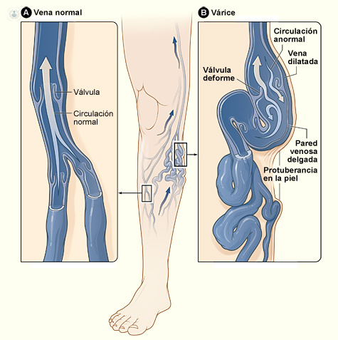
In the venous sector :
- Diagnosis of Deep or superficial venous thrombosis
- Venous Insufficiency and Varicose Veins
- Aneurysms
- Malformations
Patient preparation for echo-Doppler?
The patient, except in abdominal Doppler ultrasound, does not require fasting or other preparation, except, perhaps, to come with comfortable clothes and easy to remove and put.
Echo-Doppler Risks
As we have said, it is a non-invasive test, that is, no injections, channels, or use contrasts that can produce an allergic reaction or damage to an organ.
If the echo-doppler-color (triplex) has not been diagnosed, it will use a "eco-enhancer", venous, sugar-based, non-allergic, but it should be used with care in diabetic patients. But this test is done exceptionally.