Know the details about peritrochanteric pain or lateral pain in the hip
Written by:Pain in the side of the hip is a common symptom of consultation. The differential diagnosis must be made, by the specialist in Traumatology , between the intra-articular pain, extrarticular pain and referred pain of the lumbar spine. The lateral pain of the hip has been termed, classically, trochanteritis. Today, thanks to a better anatomical knowledge and tests such as magnetic resonance imaging (MRI), we talk about peritrocantérico pain or the trochanter pain syndrome Mayor, English "Greater Trochanteric Pain Syndrome (GTPS)". This pathology includes different alterations in the lateral region of the hip, around the major trochanter, including trochanteric bursitis, tendon lesions of the middle and lower gluteus, and hip in spring.
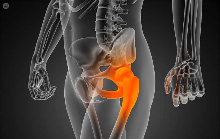
Location of major trochanter and lateral hip pain
The major trochanter is the part of the bone of the femur at the junction between the neck and the shaft. The lateral gluteus is inserted in the lateral and posterior part, and in the anterior part, the gluteus minor. These are covered by the gluteus maximus, the tensor fascia lata and the ileotibial band, which extends from the lateral side of the thigh to below the knee. Among these muscle groups the bursas are located (more frequently 3 but may be 4). The largest is located between the gluteus maximus and the tendon of the gluteus medius (trochanteric bursa), the other two, below the gluteus medius and below the gluteus maximus.
Major pathologies related to hip lateral pain and how to treat them
- Trochanteric bursitis is the inflammatory picture around the major trochanter, usually irradiated on the side of the thigh or buttock. It usually originates as a consequence of repetitive movements of hip flexion and extension, by friction between the ileotibial band and the greater trochanter. It is more common in women than in middle-aged men. It presents as pain in the lateral aspect of the hip , with pain in greater trochanter and increases with the abduction against resistance. The simple radiography is usually normal but the intra-articular pathology should be ruled out. Calcifications can be detected around the trochanter, but are non-specific.Magnetic resonance imaging (MRI) may show inflammation of the bursae and rule out tendon pathology of the gluteal musculature. Trochanteric bursitis is usually limited and may respond to rest, anti-inflammatory treatment, local ice and physical therapy. If this is not enough, infiltration with cortico-steroids and local anesthetic is usually effective in most patients. Infiltration is more effective in the short term, whereas stretching physiotherapy seems more effective in the long term. In exceptional cases surgery may be indicated, either open or by endoscopy.
- Tendonopathy of the gluteus medius and gluteus minoris are lesions of the tendons of the abductor musculature of the hip that have been equated with the rotator cuff of the shoulder. In the cases of peritrochanteric pain, due to MRI, lesions of the middle and lower buttock tendons are diagnosed, from tendinitis (inflammation) and tendinosis (degeneration) to partial ruptures or complete ruptures. These lesions are more frequent in the gluteus medius than in the minor.Although the onset of symptoms may be traumatic in origin, in most cases it is insidious and not traumatic. In addition to pain in the lateral aspect of the hip and in trochanter, the patient shows weakness to extension abduction and to external rotation with hip flexion of 90º. Pain can also be reproduced by holding the monopodal charge on the affected side for 30 seconds or more. The diagnostic test of more performance is the MRI, which allows to distinguish between partial and total ruptures , and allows to evaluate the possible fat degeneration of the musculature .Patients with gluteus tendon lesions may present a thickening of the fascia lata tensor in MRI compared to the contralateral. The initial treatment of this pathology is conservative , by means of rest, anti-inflammatory and physiotherapy , to reinforce the musculature. Surgical treatment is exceptional, in very limited cases. Repair can be done by open approach or by endoscopy.
- The hip hip or saline thigh is described as the audible and potentially painful prominence of the hip in activities requiring flexion, extension, and abduction. With the hip in extension, the ileotibial band is later located in the greater trochanter, when the hip flexes the ileotibial band slides on the trochanter. Occasionally this prominence can cause inflammation and pain. It usually occurs between the end of adolescence and the second decade of life. The X-ray is usually normal, the ultrasound, being a dynamic test, can visualize the protrusion and if there is an associated bursitis. The treatment is usually conservative , with stretching and muscle strengthening. Rarely, the treatment is surgical, with multiple techniques described, both for open surgery and for endoscopic technique.