How and When to Seal Permeable Oval Foramen (FOP)
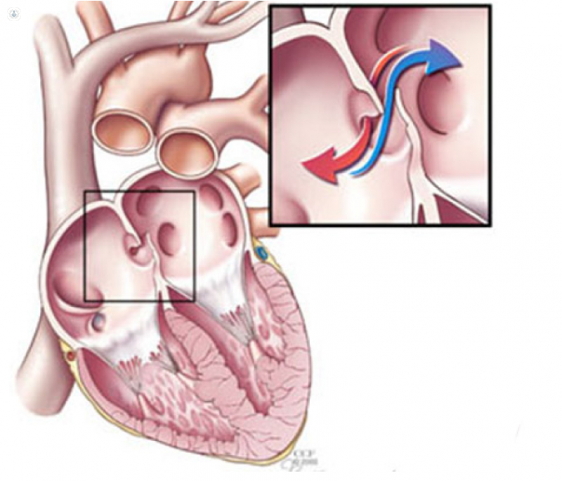
Written by:Permeable Foramen Ovale (FOP) is a communication that exists between the septum that divides the two atria of the heart.
This FOP is usually sealed by a membrane, but a failure during the embryological period may cause it to not be hermetically sealed and remain open. This circumstance occurs in approximately 25-27% of the population, and usually does not cause problems, since the pressure in the left atrium is usually greater than in the right, so it pushes the FOP membrane to the right, And there is no blood flow from the right side of the heart (low oxygenated), on the left side. The mixing of blood on the right side (venous) and left (arterial) blood can cause problems.
The causes that increase the pressure on the right side are Valsalva maneuvers, which means that intrathoracic pressure increases (lifting large weights, cough maintained, etc.).
- When FOP is closed, the passage of a small thrombus (usually from the lower limbs), migrate to the lung, which is a large reservoir that admits such elements, without major symptoms.
- But when pressure on the right side increases, for whatever reason, and the FOP membrane is open, a small thrombus can pass from the right to the left side and migrate to the carotid arteries, causing a stroke.
- One circumstance that is very prone to these accidents is the deep diving with compressed air. If a correct decompression is not performed, the nitrogen accumulated in the tissues and that diffuses very slowly, migrates in large bubbles to the venous system. As during the dive several maneuvers are performed that increase the right pressure (Valsalva), if you also have the bad fortune to have the FOP permeable, it is easy to produce a stroke.
- Therefore, before performing dives, an Echocardiogram should be performed for safety, and if there is FOP, make another test called Transesophageal Echocardiogram with sonic contrast, to determine if there is a right-left passage, and if it is in the amount of> 15-20 microbubbles. In that case, it is advisable to limit diving to shallow depth and without requiring decompression or to close the FOP definitively.
And what is done in case there is in any case an FOP with right-left short circuit?
Much has been discussed. There are anatomical variables that favor the occurrence of strokes (FOP size, bubble passage number, exaggerated growth of Eustachian Valve (intracardiac structure facilitating the passage of venous blood to the left atrium, if there is FOP) .
How and when to seal the FOP
In any case, before a young person - a diver or not - who presents a Cryptogenic Stroke (that happens after excluding any Cardiac, Neurological or Hematological causes) that favors it, the opinion is to close it percutaneously. This is done through a cardiac catheterization and is closed by implanting a device in the form of a diabolo, which closes the FOP.
However, while there are small controversies about this type of action, it is the usual practice in most of Western Europe and Anglo-Saxon countries.
For more information consult a cardiologist .