All you need to know about the stones in the urinary tract
Written by:Urolithiasis is the formation of stones within the urinary tract by deposition of molecules found in the urine diluted, in susceptible individuals.
In Spain it is estimated that more than 5% of the population suffers from urolithiasis, although more recent studies raise this figure to about 10%. This disease is 1.6 times more common in men than in women.
Levels where urolithiasis is formed
Urolithiasis stays at different levels of the urinary system:
- nephrolithiasis: is formed inside the kidney and is the most common.
- ureteral calculi: usually migrated from the kidney towards the bladder, rarely form in the ureter. These stones are usually not greater than 1 cm and often obstruct the passage of urine.
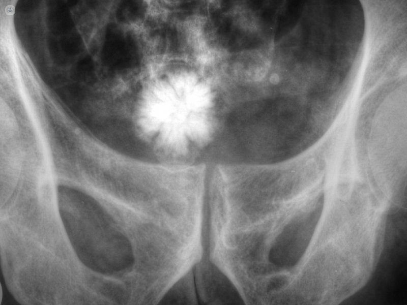
- bladder stones: is formed in the bladder and affects men over 50 years.
- Prostatic calculi: it develops in the prostate.
Causes of urolithiasis
The causes that promote gallstone formation may be varied, but often attend more than one in the same person, whether by external factors such as the patient. Of external factors, the most prominent are:
- Climate: in the hottest months of the year, episodes of urolithiasis because of the lack of hydration are more frequent. It has also been proven that workers in situations of heat and dehydration and sedentary work positions have an increased frequency of urolithiasis.
- Hydration: Insufficient water intake tank favors salts in the urinary tract and, therefore, the development of lithiasis. As for the type of water, it has not been shown that there is more risk of developing stones in water more or less calcium content.
- Diet: lack of calcium in the diet can promote the formation of stones, as this increases the excretion of calcium oxalate in the urine and thus may appear litiasis. On the other hand, excess salt diets also favor the onset of this disease.
- Drugs: some diuretics, corticosteroids and antiretrovirals can also cause stones.
On the other hand, the patient's conditions also influence the origin of urolithiasis:
- Age: urolithiasis is less frequent until age 20, while between 40 and 60 years for men and between 50 and 60 years in women is more prevalent.
- Family history: patients with relatives who have had stones have three times the risk of suffering also.
- metabolic syndrome and obesity: diabetes and insulin resistance favor the appearance of stones.
- Hypercalcemia or hypercalciuria: disorder that causes increased calcium from any cause.
- Hyperthyroidism.
- Hyperuricemia: favors the formation of calculi of uric acid and calcium oxalate.
- intestinal malabsorption: increases the absorption of oxalate.
- Metabolic disorders: primary hyperoxaluria, cystinuria, cystic fibrosis or renal tubular acidosis.
- morphological alterations in the urinary tract: stenosis of ureteropelvic junction, caliceal diverticula, megaureter, sponge kidney, prostate hyperplasia obstruction or urethral stricture (the latter may favor bladder stones).
- Recurrent infections: affects more carriers ureteral catheters and urinary catheters nephrostomies, especially in women.
- Immobilization: patients with severe mobility limitations are more likely to form stones in the urinary system.
Symptoms of urolithiasis
Symptoms depend on the location of the stones, the size, causing obstruction in the urinary tract and superinfection they can generate.
Frequently, the stones do not cause symptoms and are discovered by chance in a radiology test (x-ray or ultrasound). Now when a stone obstructs the urinary tract, then the patient feels pain.
This pain, called renal colic is caused by the accumulation of urine above the stone, which distends the urinary tract. Normally, renal colic pain is intense, alternating periods of increased pain relief with other apparent. It is accompanied by nausea and vomiting and sometimes constipation reflects intestinal paralysis.
Bladder calculi do not cause obstruction in the urinary tract, but are formed by chronic to the emptying of the bladder obstruction, and are usually accompanied by irritability, frequent urination, dull ache in the lower abdomen, pain after urination or bleeding in the urine.
Consequences of urolithiasis
Obstruction of the urinary tract increases the pressure within it and progressively damages the affected kidney. The kidney undergoes a series of changes which, after a variable time (according to the previous situation and if the obstruction is total or partial), ends up override function temporarily, if unclog, or permanently, if it persists the problem.
In a person with two functional kidneys, blockage of one of them does not have to alter the overall renal function, as the other body replaces obstructed work. However, it is common for people with large body size or muscle mass, especially males, develop a mild renal insufficiency by renal colic with complete obstruction. This renal failure is reversible and generally has no effect.
On the other hand, a person with a single functioning kidney may develop anuria if this is blocked, ie can stop the excretion of urine. If the situation persists, a rapidly progressive renal failure can be deadly within days if the blockage is not resolved or no recourse to dialysis (removal of harmful substances retained) set. This can occur with renal colic and painlessly.
People with obstructive partially functioning kidney stones and one can develop kidney failure by maintaining a seemingly normal urine output, although it is a less common situation.
Complications of urolithiasis
There are certain groups of people are more likely to have serious repercussions if they have urolithiasis:
• People with disabilities
• monorenal (people with a functional kidney)
• Patients with increased risk of infection
• Pregnant
A complication of stone formation is hydronephrosis, dilation consisting urinary tract. It is easily diagnosed by ultrasound and can be classified into four grades:
• Grade I: mild dilatation without affecting the shape of the calyces. • Grade II: dilated calyces without loss of renal mass
• Grade III: Severe dilation deformation of chalices, without thinning of renal tissue.
• Grade IV: dilation severe loss of kidney tissue.
Sometimes the stones, even large intrarenal size, go unnoticed and can be diagnosed in an advanced situation in which the kidney has irretrievably lost its function hydronephrosis grade IV or a situation in which the kidney is no longer dilated, but has lost volume (renal atrophy).
Another complication of gallstone is the gallstone or complicated pyelonephritis, infection of the urinary tract that affects the kidney and urologic constitutes an emergency as it can lead to sepsis (severe infection and life - threatening). In this situation, it is recommended to some patients placed in the urinary tract system that will avoid the obstruction caused by gallstone, usually an internal or external catheter or nephrostomy (derive the course of urine from the kidney to the skin).
Treatment of urolithiasis
In the pharmacological field, there is no effective specific treatment for most stones that have already formed. Only lithiasis composed of uric acid have medical treatment consisting of drugs that increase urine pH, ie, reduce their acidity, and are capable of dissected.
Once formed the pharmacologically untreatable stones, we must resort to surgery to remove it completely. It is advisable to visit a specialist in Urology to choose surgical treatment necessary depending on the type of gallstone suffering:
- Extracorporeal shock wave lithotripsy: A procedure that involves the application of shock waves (pressure waves) at the closest point to the skin of urolithiasis. These waves pass through the tissues to reach the stones and fragmented, without surgery. In principle, the fragments are subsequently excreted in the urine. It is not recommended for patients with large stones, since a large amount of fragments would be generated and therefore would be difficult to pass and certainly require multiple sessions, which can last for months.
- Percutaneous nephrolithotomy: surgery, usually under general anesthesia, which is to access the kidney through a small hole in the side, through which an endoscope is inserted (to see inside the kidney) and a power source (one laser, for example) to fragment the stones, removing debris later. Unlike lithotripsy, it is advisable for patients with large stones, as the resolution rate is higher and the procedure is faster.
- Ureteroscopy: surgical procedure usually performed under spinal anesthesia (in the intrathecal space), sometimes using local anesthesia, which involves inserting a long, thin endoscope through the urethra to access the place where the stones and fragment it by laser or another energy source. It is used to treat ureteral stones but can also deal with kidney stones. The advantage of this technique is endoscopic having a high resolution rate and generally requires only one procedure.
- Open surgery is an exceptional surgical treatment, only for very complex cases where it is necessary to remove a portion of the kidney or urinary tract remodel. It may also be an option in the rare circumstances in which it has not managed to solve the stones by the methods described above.
- bladder stones: are usually treated endoscopically, through the urethra. If they are associated with an obstructive problem (prostatic hyperplasia or urethral stenosis), also it is solved sometimes by open surgery (in the case of large prostates and large stones and / or multiple).
- Prostatic calculi: is not clinically relevant and does not need to treat them.
Prevention of urolithiasis
For patients with kidney stones, you need to take some preventive measures, regardless of the level of risk:
- Drink between 2.5 and 3 liters of fluid a day
- Ingesting liquid form distributed throughout the day
- balanced diet with normal calcium content, low salt, and adequate amount of fruits and vegetables
- Maintain an appropriate weight
- Moderate exercise
- Avoid high consumption of foods rich in oxalate (spinach, beets, chard, cocoa powder, nuts, etc.)
- Avoid high consumption of foods rich in animal protein (in patients at risk of gallstone formation of uric acid)