Types of retinal detachment
Written by:Retinal detachment occurs when the retina, a light sensitive membrane is separated from its supporting layers. Specialists in Ophthalmology claim that these layers are in contact but not attached, so there may be situations that cause the retina to detach from this support are known as pigmented epithelium layer.
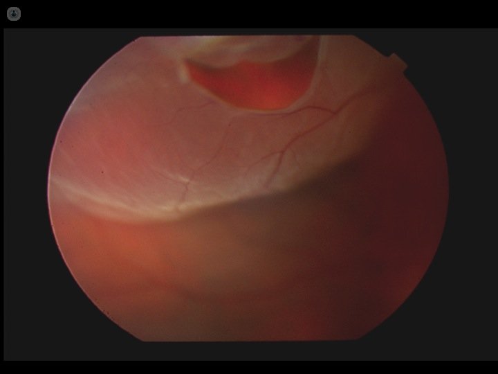
When a retinal detachment occurs, the retina degenerates and deteriorates, because the pigment epithelium is the tissue responsible for feeding, caring and deliver oxygen to the retina. If a retinal detachment occurs, these actions are not carried out and, therefore, the retina loses its function of perceiving the light is focused from outside. This causes a progressive loss of vision that, if not treated in time, can lead to blindness.
Types of retinal detachment
The most common retinal detachment is regmatóneo, when filtered fluid from the vitreous cavity to virtual subretinal space through a break in the retina. Another type of detachment is the origin Tractional. This occurs due to traction towards the center of the eye and fibrotic membrane proliferations which are attached to the retina, as in the case of people with advanced diabetes. Finally, it gives serous exudation of fluid between the two layers due to inflammation, tumors or other various reasons.
Treatment for retinal detachment
The rhegmatogenous retinal detachment or tensile type type is treated by surgery. These types of release are produced in most cases. Retinal detachment requires serous its root cause, which may be by surgery or with drugs or radiation therapy, and others concerned.
There are different types of surgery performed according to each case, but currently the most widely used technique is by microsurgery via pars plana posterior vitrectomy.