Improvements in Mammography Technique
Written by:The objective of mammography is the radiological study of mammary pathology. It has to be performed from the age of 40 on an annual basis.
The problem with mammography is that it is uncomfortable for most women, and for a minority it can even be painful; everything depends on the threshold of the pain of each one, its predisposition and the fear of the final result.
Advantages of the Senographe Pristina system in mammography
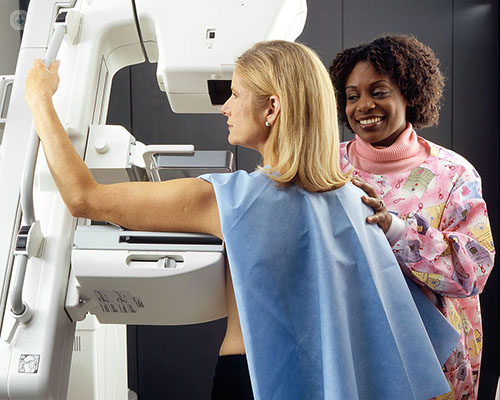
Mammography is less troublesome when performed during the first nine days after the onset of menstruation. The main factor that propitiates is the compression of the breast for a few seconds, aiming to achieve a clear image. In this way, in addition, the gland immobilizes and decreases the radiation to be used.
However, with the new system of self-control of the mammary pressure and of the mechanisms of relaxation that we use in our center, the annoyances are smaller.
This is Senographe Pristina, a technology that allows the patient herself to control the compression of the breast, which makes it different from traditional mammography, which presses the chest automatically.
With this new tomosynthesis system you can get a very clear and detailed 3D breast image, with radiation exposure similar to that of a conventional mammogram and avoiding repetitions of the test (necessary when the deficient shots are deficient). In this way, in some cases the patient receives less radiation, because it undergoes less tests.
Advantages of the ABUS Invenia system in mammography
As we did with Senographe, we were the first to implant in Spain an integral breast diagnostic system that includes an automatic ultrasound (Abus: Automatic Breast Ultrasound), capable of performing diagnostic tests in just 15 minutes.
Automatic breast ultrasound is an ultrasound technology that does not emit radiation and is especially suitable for testing in dense breasts, that is, in those in which there is a greater volume of breast tissue than fat. In this type of breast, up to one-third of the tumors may go unnoticed by traditional mammography because the breast tissue can mask the suspicious lump, seeing the same color in the image. In contrast, using automatic ultrasound clearly differentiates cancer from breast tissue.
For more information consult a radiologist .