How mediastinal disease diagnosed?
Written by:Diagnostic imaging tests are very useful to detect diseases of the mediastinum. Specialists in thoracic surgery claim to be a non-invasive and completely painless for viewing images of organs and identify any abnormalities in the patient's body.
 In the case of the mediastinum, most disease have no previous symptoms, so sometimes the chest radiography may show injuries and traumas that were previously ignored, whose diagnosis will be complete only after some imaging tests.
What is the mediastinum?
The mediastinum is the anatomical compartment of the chest that lies between the two pleural cavities, and contains all the thoracic organs except the lungs. Very often in the mediastinum they can settle masses (tumor or cyst) or infections (acute or chronic). These masses can represent different diseases, among which are thymomas, lymphomas, intrathoracic goiters, teratomas or neurofibromas.
What to do in case of injury or trauma?
After a discovery of this kind, it is advisable to conduct additional studies and specialist conducts an evaluation of the clinical history of the patient, considering his age, symptoms, location and size of the mass found.
The presumptive diagnosis is made after a careful imaging tests, which provide an optimal definition of the lesions in the mediastinum and its relationship with neighboring organs. A needle biopsy guided by CT or surgical establishes the definitive diagnosis.
Major diagnostic imaging tests for diseases of the mediastinum
- TC or Scanner: Also called CAT or CT scan is the test most commonly used image diagnosis of mediastinal tumors. This is a very complex machine, consisting of a bed in which the patient is placed, and X-ray tube turning rapidly to acquire information of the entire volume of the patient's body.
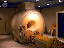
- Magnetic resonance imaging (MRI): An MRI is an imaging test that uses magnets and radio waves to create powerful images within the body without using X-rays or radiation. It is indicated to differentiate between vascular mediastinal tumors or bronchial. It is used to assess tumor invasion heart, great vessels, chest wall and spine. It is also more accurate than CT in distinguishing cysts from solid tumors.
- PET: Positron emission tomography (PET) differs from the other above named techniques that can show chemical and physiological changes related to metabolism, and therefore, these images can show abnormalities and changes in the tissues. First, a radiopharmaceutical injected into the patient, called isotope. After an amount of X-rays through the patient's body helps determine the details of cellular metabolism. PET is useful for predicting the degree of malignancy of tumors, and to assess the presence of residual disease after chemotherapy.
- Esophagogram: A study that involves taking X-rays of the patient in various positions for imaging the esophagus, in order to determine changes in the normal anatomy. It is sometimes used to assess mediastinal lesions affecting it.
- Digital Angiography is a test whose function is to study the circulatory vessels that are not visible by conventional radiological examinations. It is used in some neurogenic tumors with intraspinal extension, and can replace the RM
- Gamma-imaging studies: There are three types of gamma-imaging studies:
- Scintigraphy with iodine, used in the study of endotorácico goiter or thyroid carcinoma metastases.
- Gallium, which is used to evaluate acute and chronic mediastinal infections.
- Technetium scintigraphy can be used to identify gastric mucosa in the visceral compartment Neurenteric cysts.